

Diastolic collapse of the right heart chambers suggests hemodynamic compromise in the presence of pericardial effusion. Right atrial collapse is believed to occur at an earlier stage of hemodynamic deterioration than right ventricular collapse. (David Wrisley, MD, Syracuse, New York).
What causes right atrial collapse?
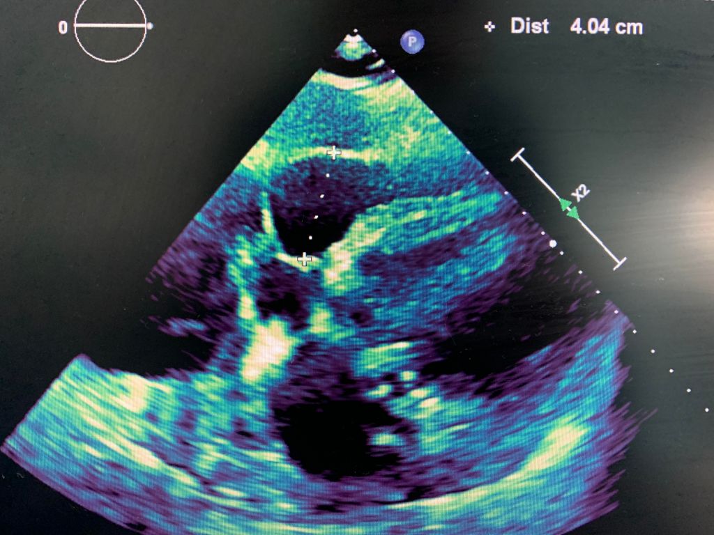
A large pericardial effusion. It occurs during early to mid diastole and collapse of the right atrium occurs in late diastole. 3 These events are seen echocardiographically when equalization of in trapericardial and intracardiac pressures occurs. (David Wrisley).
Case Study:
A 78 y/o Female presents for complete echocardiogram with a history of dyspnea on exertion. Patient is on 4 L of oxygen, unable to breathe laying supine. Breathing gets better as she sits up. Below are just a few images of her echo.
It was agreed by the physicians that the patient had a large pericardial effusion and her right atrium was collapsing. Further treatment to be determined.
Tips when doing echocardiogram:
- PLAX view: measure any effusion you see
- PSAX view: use M-Mode through the Mitral Valve to show ventricular wall motion; reduce the sweep speed to 100 mm/s and compare the Right Ventricualr wall motion to the Mitral Valve motion.
- Apical 4: perform MV and TV respiratory variation Doppler by placing the PW gate at the tips of MV and TV (make sure you annotate each valve separately). Decrease the sweep speed to 25 mm/s. Mark any changes in mitral valve peak E velocity – if the change is greater than 30% then we may have possibility of tamponade). Mitral valve inflow decreases with inspiration and increases with inspiration; however, tricuspid valve inflow increases with inspiration and decreases with expiration.
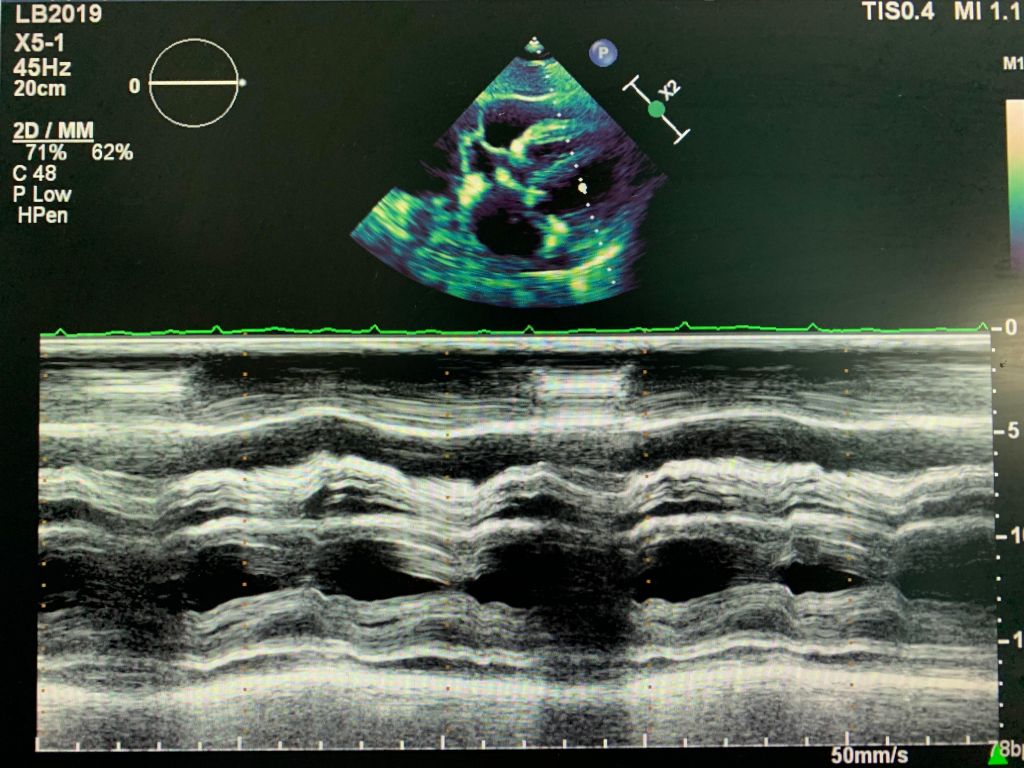
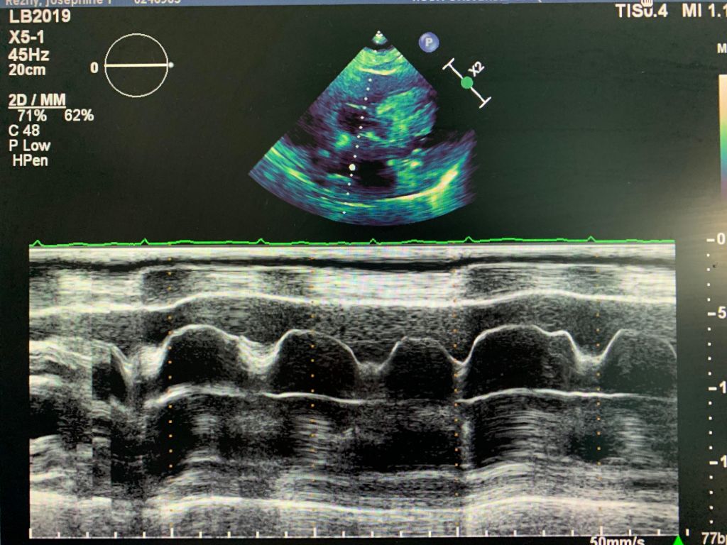
- SUB4: perform M-mode through the ventricular walls and decrease the sweep speed to 100mm/s to assess the wall motion.
Below pictures are taken from the SUB4 view only, as the patient’s respiratory variation didn’t show any changes.
As always, work has been cited:
Marked Diastolic Collapse of the Right Atrium Without Hemodynamic Compromise Caused by a Large Pleural Effusion
DOI:https://doi.org/10.1016/S0894-7317(14)80424-0
https://www.onlinejase.com/article/S0894-7317(14)80424-0/pdf
