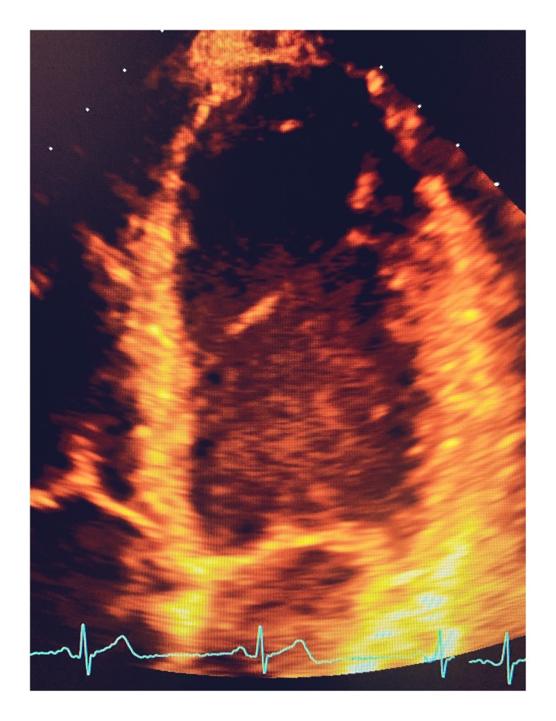
False Tendons
Left ventricular false tendons are fibrous or fibromuscular bands that stretch across the left ventricle from the septum to the free wall. They can also tether to a papillary muscle, but unlike the chordae tendineae, do not connect to the mitral leaflets. They are anatomic variants that should not be mistaken for abnormalities such as tumors, subaortic membranes, thrombus borders, septal hypertrophy. They have been noted in patients with murmurs and arrhythmias.
Left ventricular (LV) false tendons are chordlike structures that traverse the LV cavity. They attach to the septum, to the papillary muscles, or to the free wall of the ventricle but not to the mitral valve. They are found in approximately half of human hearts examined at autopsy. Although it has been more than 100 years since their initial description, the functional significance of these structures remains largely unexplored. It has been suggested that they retard LV remodeling by tethering the walls to which they are attached, but there are few data to substantiate this. Some studies have suggested that false tendons reduce the severity of functional mitral regurgitation by stabilizing the position of the papillary muscles as the left ventricle enlarges. LV false tendons may also have deleterious effects and have been implicated in promoting membrane formation in discrete subaortic stenosis.
JASE online https://www.onlinejase.com/article/S0894-7317(13)00176-4/abstract
Credit: http://rwjms1.umdnj.edu/shindler/false_tendon.html